
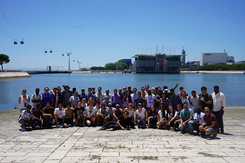
We are still trying to understand exactly how the human brain works.
In conditions such as epilepsy, in which people suffer from sudden attacks (which can imply a loss of consciousness) what happens in the human brain to cause these events is still relatively unknown. The goal of better understanding these factors to better help people suffering from diseases like these is what led researcher Rodolfo Abreu, guided by Professor Patrícia Figueiredo, to want to use the techniques of the Electroencephalogram (EEG) together with Magnetic Resonance Imaging (MRI) and combine them to have a clearer notion of what is causing these faults.
“We took the EEG equipment to a hospital, put everything inside the MRI scanner and checked the images simultaneously. Afterwards, I processed the data offline, to try to complement and take advantage of the best things in both techniques. The EEG has a very good temporal resolution – you can see things that happen very fast in the electrical activity of the brain – but in terms of spatial location it’s not very good. The MRI is, in some ways, the exact opposite.”
Rodolfo explained how epileptic events can be either isolated or what is commonly known as ‘epileptic attacks’ and how both of those are very quick to happen and often fade just as quickly. “These events can very easily be identified with the EEG when they are happening but you would need the MRI to know where in the brain, so this combination of the two can be very fruitful. Ultimately we would want to know which brain region is responsible for generating that abnormal brain activity so it becomes an option for the patient to have it removed.”
Working with both techniques offers very specific problems. If separately each exam can have its own difficulties – if you blink that might come up as an artefact because of the electrical activity generated or how our own heartbeat can generate fluctuations in the analysis – then when trying to simultaneously acquire data from different exams it can be an even more difficult task. “Usually when you use an EEG at an MRI machine the EEG signal can be completely destroyed, even though that, thankfully, that’s possible to correct. During my PhD, I worked on trying to find methods that manage to remove those artefacts as much as possible without also removing what is important. The most complicated part is the ‘translation’ from one technique to another. If you want to combine two completely different things you cannot just fuse them together.”
From one particular condition to something even bigger.
Different regions of the brain have anatomical connections, but regions can also be connected by function. And those connections can change according to your cognitive state or activity. Even in a resting state, you have particular neural networks if you are tired, or listening to background noise. “We wondered, what if, even as a resting state, there could be specific networks associated with the disease, like a pathological network that is always there.” This became the next stage of Rodolfo’s work: to study the dynamics of these variations between networks.
“Typically, you divide the brain into regions and then try to calculate the force of the connection between those regions. This would get you a value for each connection. But to realize if this varies in time you need to take the data but calculate the value for only a period of time. This gives you different instances and allows you to realize the differences in connection. We then can use methods of pattern recognition to be able to recognize specific representations of what is happening in these areas of the brain. Those states can be further analyzed to figure out if some might be associated with the disease, either contributing directly or being associated in some way. Associated dynamics and long-term variations are what allows to, together with the clinical input, take conclusions from the brain activity of the patient, even if they are in a resting state.”
Understanding these factors could lead to fruitful options, like knowing how functional activity can alter anatomical activity, something of great importance to increase safety when there’s the possibility to detach the problematic area.
Knowing that these techniques see the same thing although in different ways and knowing that there’s a way to create a bridge between techniques means having a way to more safely comprehend what happens in the brain, not only with epilepsy. “These tools are transportable to other issues because it offers tools that can be used whenever the aim is to cross-check information from these two different exams.”
New projects are coming up that can use these tools to help even more people. Patricia Figueiredo explained how a new project aiming to study the effects of migraines is using some of the same tools on different goals. The current project is undertaken not only with the medical staff, but also includes neuropsychologists working with the patients to assure the collaboration in the necessary stages. This includes two PhD candidates who are co-oriented both by ISR and Faculdade de Medicina of Universidade de Lisboa.
“We want to further understand the disease through the complete the migraine cycle, so not only when the patient is experiencing pain, but also in other more restful states. This is only made possible because of a tight collaboration with the physicians and therapists involved in the care of the patient, that allows us to be able to get the data from these exams at times other than those of emergency. It’s exciting to be able to think in such a rounded way.”


